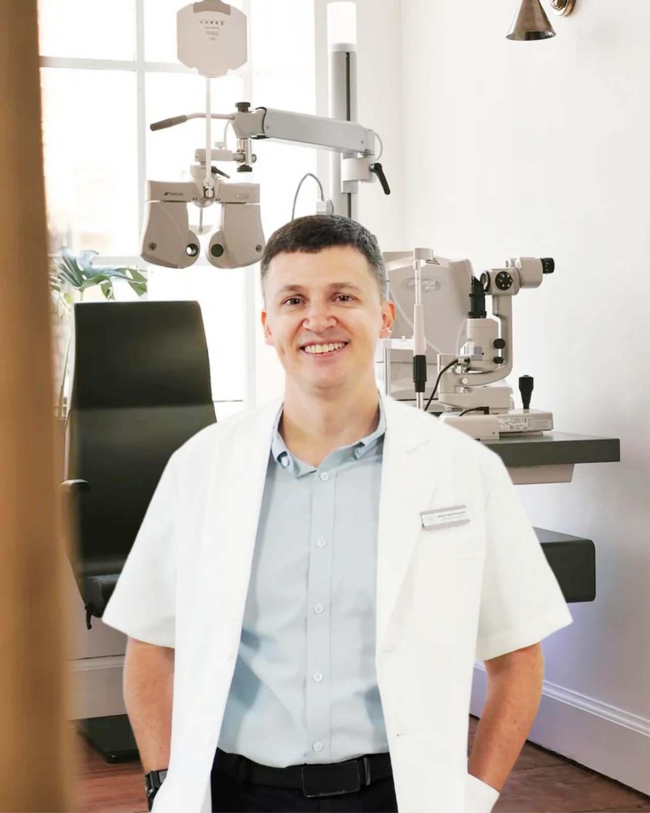
Lead Ophthalmologist · Cataract and Retina Surgeon
Dr. JD Ferwerda, MD
Dr. JD Ferwerda, MD: Dutch-trained ophthalmologist specialising in cataracts, lens exchange, retinal disease, glaucoma, and AMD.
When a cataract starts affecting your daily life — driving, reading, recognising the faces across the dinner table — surgery removes the clouded natural lens and replaces it with a clear artificial one.

Cataract surgery is the removal of the eye's clouded natural lens and the implantation of a clear, permanent intraocular lens (IOL) in its place. The natural lens sits just behind the iris in a thin elastic envelope called the capsular bag. Over time the protein structure of that lens stiffens and yellows; that is what a cataract is. Once it begins to interfere with everyday tasks, surgical replacement is the only meaningful treatment — no drops, glasses, or supplements reverse a true cataract.
The good news is that the procedure has matured into one of the most refined operations in medicine. It is small-incision, day-case, and performed under topical anaesthesia in most patients. Modern IOLs do more than restore clarity — they correct astigmatism, address presbyopia, and let many patients reduce or eliminate their reliance on glasses. The choice of lens, and the timing of surgery, is what the consultation is for.
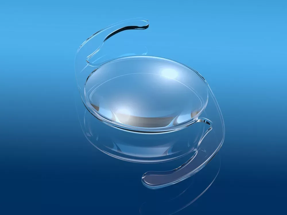
The surgery begins with a self-sealing incision of around 2.2 to 2.8 millimetres at the edge of the cornea. A circular opening is made in the front of the capsular bag, leaving the rest of the bag intact to hold the new lens. A fine ultrasound probe is then introduced through the incision; it vibrates at high frequency to gently fragment the cataractous lens.
Those fragments are aspirated out of the eye through the same probe, leaving the empty capsular bag in place. A folded artificial lens is then loaded into an injector and slid through the incision; once inside the bag it unfolds, centres itself, and settles. The incision is typically too small to require stitches and seals on its own as the eye rehydrates.
The choice of IOL is what shapes the visual outcome. Different lens designs solve different problems — sharp distance vision, correction of astigmatism, freedom from reading glasses — and the recommendation depends on your eye anatomy, your dominant tasks, and your tolerance for the small visual trade-offs of premium-optic designs.
The right time to operate is when the cataract is interfering with the life you want to lead — not before, and not so far after that the surgery becomes harder than it needs to be.

A cataract that is making daily activities harder — driving at night, reading menus, recognising faces, following a screen

A glasses prescription that has shifted noticeably in the past year or two, suggesting nuclear sclerosis

Glare, halos, or starbursts around lights that didn't bother you a year ago

Colours looking washed out, yellowed, or dim — especially noticeable when one eye is more affected than the other
Not the right fit if: the cataract is present on examination but not yet affecting how you live. Monitoring at six- or twelve-month intervals is the right answer. Surgery is a tool used at the right time — not the moment a cataract is named.
Cataract surgery itself takes an afternoon. Everything around it — the planning, the IOL choice, the year of follow-up — happens with the same surgeon at the clinic.
Optical biometry, IOL power calculations, corneal topography, dry-eye check, and a slit-lamp examination. Dr. JD walks you through findings and IOL options the same day.
You arrive at our partner facility. Drops dilate the pupil and numb the eye. The procedure itself takes about 15–30 minutes; you are in the theatre suite for around an hour in total.
You go home a few hours after surgery with prescribed eye drops, a clear shield to wear overnight, and a written aftercare plan. Most patients sleep, and notice clearer vision the following morning.
Day 1, Week 1, and Month 1 reviews back at the clinic, with the same doctor and the same chart. A one-year check confirms long-term stability.
Most patients are seeing well within a couple of days. The drops and the activity restrictions are what protect that result through the first month.
Slightly blurred or watery vision is normal as the eye settles. Expect mild grittiness or scratchiness rather than pain. Wear the protective shield while sleeping. Begin the prescribed drops on schedule.
No heavy lifting, no swimming, no rubbing the eye. Sunglasses outdoors. Most desk work resumes within 2–3 days. Vision sharpens noticeably across the week.
Tapering schedule of anti-inflammatory and antibiotic drops; vision continues to refine as the eye stabilises. Avoid dusty or contaminated water until cleared at the Month-1 review.
Long-term review at twelve months. The IOL itself is permanent and stable; occasional clouding of the posterior capsule is treated in clinic with a brief YAG laser procedure if it ever appears.
Dr. JD Ferwerda, MD: Dutch-trained ophthalmologist specialising in cataracts, lens exchange, retinal disease, glaucoma, and AMD.