Have you suddenly noticed your central vision becoming blurry, distorted, or a dark spot appearing right in the middle of your sight? Don’t ignore it, as this could be a sign of a macular hole – a serious condition that directly affects the macula, a small but critically important area responsible for the sharp vision in your eye. A macular hole, though painless, significantly impairs your ability to read, drive, recognize faces, and see fine details.
To protect your precious central vision, it is crucial to understand what a macular hole is, its causes, early macular hole symptoms to recognize, and effective macular hole treatment methods. This article will serve as a comprehensive guide, providing you with essential information concisely, succinctly, and in the easiest-to-understand manner.
What is the Macula and Why is Central Vision So Important?
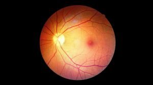
Before delving into macular holes, we first need to understand the macula. The macula is a small, oval-shaped area located precisely in the center of the retina – the light-sensitive tissue at the back of the eyeball. Although it occupies only a very small portion of the retinal area, the macula plays a pivotal role, being responsible for:
- Sharp central vision: Ensuring the ability to see the smallest details clearly.
- Color vision: Helping the eyes distinguish various shades and hues.
- Ability to perform high-acuity activities: Such as reading, safe driving, threading a needle, or accurately recognizing faces.
In essence, the macula is considered the “heart” of your vision, playing a crucial role in helping you interact with the surrounding world as clearly as possible.
>> Age-Related Macular Degeneration (AMD): Causes, Symptoms, and Prevention
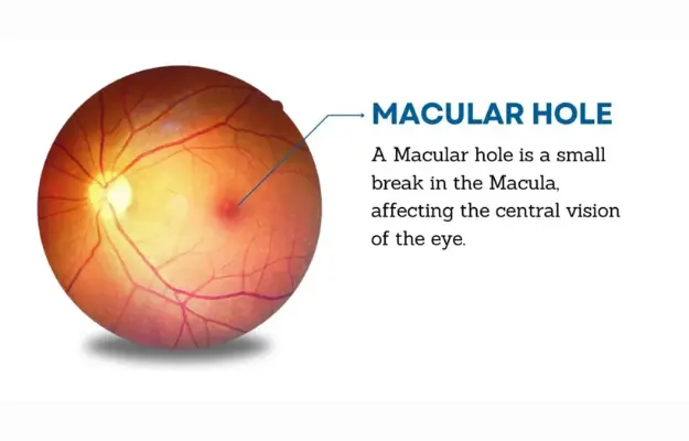
What is a Macular Hole?
A Macular Hole is a small tear or perforation that appears right in the macula – the central area of the retina. This condition causes the images received by the macula to become distorted, blurred, or completely lost, leading to a significant decrease in central vision.
Nature of the macular hole condition:
- Painless: Despite being a tear, a macular hole typically does not cause pain in the eye.
- Directly affects central vision: Leading to macular hole symptoms such as blurred vision, distorted images, or the appearance of a blind spot directly in the center of your field of view. This differs from peripheral vision (surrounding vision), which is usually unaffected.
- Often occurs in one eye: However, about 10-15% of patients have a risk of developing a macular hole in the other eye in the future.
Classification of macular holes by stage:
The stages of a macular hole are crucial for determining the prognosis and selecting the appropriate macular hole treatment:
- Stage 1 (Impending macular hole): There is only a partial separation of the macula’s layers.
- Stage 2 (Partial-thickness macular hole): A small hole that has not yet penetrated all layers of the macula.
- Stage 3 (Full-thickness macular hole): A hole that penetrates all layers of the macula.
- Stage 4 (Full-thickness macular hole with complete posterior vitreous detachment): The hole is fully formed, and the vitreous has completely detached from the retina.
Causes of Macular Hole: Why Does the Macula Tear?
The primary cause of a macular hole often stems from the natural aging process of the eye, particularly changes in the vitreous humor – the clear gel that fills the eyeball.
- Vitreomacular Traction (VMT): This is the most common cause. As we age, the vitreous naturally shrinks and pulls away from the retinal surface. Typically, this process occurs smoothly. However, if the vitreous remains firmly attached to the macula and pulls strongly, it can stretch and ultimately create a small hole directly in the macula.
- Eye trauma: A strong blow or severe injury to the eye can directly cause a macular tear.
- High myopia (nearsightedness): People with severe nearsightedness typically have longer eyeballs than normal, causing the retina to be stretched and thinned, thereby increasing the risk of macular hole formation.
- Retinal detachment: Although not a direct cause, retinal detachment can increase the risk of developing complications at the macula.
- Other eye conditions: Certain conditions such as diabetic macular edema, chronic uveitis, or prolonged use of corticosteroids can also increase the risk.
Age is the leading factor, with most macular hole cases diagnosed in individuals over 60 years old.
>> Common Retinal Diseases and Effective Prevention Methods
Signs and Symptoms of Macular Hole
A macular hole usually does not cause pain, but visual macular hole signs and symptoms will become apparent, directly impacting your ability to see clearly. Early recognition of these macular hole symptoms is extremely important for timely intervention:
- Blurred or distorted central vision: This is the most common macular hole symptom. Images directly in the center of your vision may become blurry, wavy, or distorted. Straight lines might appear curved or rippled (this is a characteristic sign often checked with an Amsler grid).
- Difficulty reading, seeing details: Due to affected central vision, you will experience significant difficulty reading small print, threading a needle, or seeing intricate details.
- Appearance of a dark spot or blind spot in the center of vision: As the macular hole enlarges, you might notice a dark area or a gap (blind spot) right in the middle of your visual field.
- Difficulty recognizing faces: Due to impaired central vision, recognizing other people’s faces becomes more challenging.
- Double vision (diplopia) in one eye: Although less common, some individuals may experience double vision when looking with only the affected eye.
If you notice any of the above macular hole signs, especially sudden changes in central vision, see an ophthalmologist immediately for accurate examination and diagnosis. Early detection significantly increases the likelihood of successful macular hole treatment and maximum vision preservation.
Is a Macular Hole Dangerous? When to See a Doctor?
A macular hole is a serious condition for central vision. Although it does not directly cause complete blindness (as peripheral vision usually remains), it can significantly impair the quality of life, hindering daily activities that require detailed vision.
- Severity: If macular hole treatment is not sought promptly, the tear can continue to enlarge, leading to permanent and irreversible central vision loss.
- Not an immediate emergency: Unless there are sudden and severe symptoms such as intense pain accompanied by acute vision loss (which is very rare with a simple macular hole), a macular hole is not a “minutes-matter” emergency like retinal detachment or acute glaucoma. However, delaying treatment can reduce the chances of later vision recovery.
When to see an ophthalmologist:
- Immediately: If you suddenly experience macular hole symptoms such as abrupt central blurry vision, severe visual distortion, or a large dark spot directly in the middle of your vision.
- Within a few days: If the aforementioned macular hole symptoms develop gradually or if you have noticed them for some time but haven’t yet been examined.
Remember, timely diagnosis and macular hole treatment are key to achieving the best possible visual outcome.
Diagnosing Macular Hole: Modern Methods
To accurately diagnose a macular hole, an ophthalmologist will utilize specialized examination methods and imaging techniques to thoroughly evaluate the condition of the macula and retina:
- Dilated fundus examination: The doctor will instill dilating eye drops and use a specialized ophthalmoscope to directly observe the retina and macula. This is the initial step to detect a macular hole and other abnormalities.
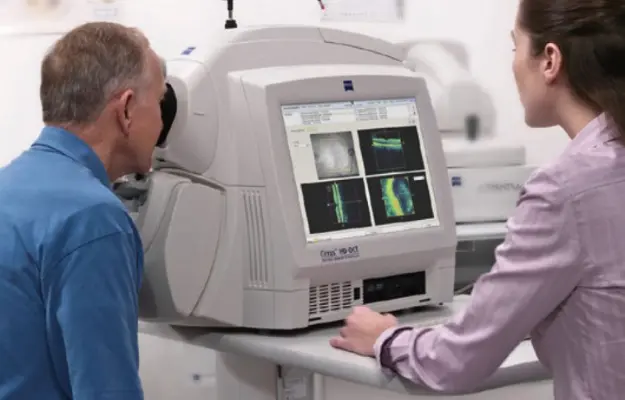
- Optical Coherence Tomography (OCT): This is the most precise and crucial method for macular hole diagnosis. OCT uses light waves to create high-resolution cross-sectional images of the retina. This imaging allows the doctor to clearly visualize the macular structure, identify the presence, size, shape, and stage of the macular hole, as well as the extent of vitreous traction if present. OCT is also highly beneficial for monitoring disease progression and evaluating post-surgical outcomes.
- Fluorescein Angiography (FA): Although less commonly used directly for macular hole diagnosis, FA may be performed to rule out other retinal conditions with similar symptoms or to assess the condition of blood vessels around the macula.
- Vision and visual field test: The doctor will test central vision and may perform a visual field test to evaluate the extent to which the macular hole affects your overall vision.
Based on the results of these tests, the doctor will provide an accurate diagnosis and recommend the most suitable macular hole treatment plan.
Macular Hole Treatment: Surgery and Other Options
In most cases, the most effective macular hole treatment is surgery. The goal of surgery is to close the macular hole, thereby helping to improve central vision.
- Vitrectomy surgery: This is the primary surgical technique used to treat a macular hole.
- Procedure: The surgeon will make a few very small incisions in the eye. Microscopic instruments are inserted to remove the vitreous gel that has pulled on or caused the macular tear. Subsequently, a gas bubble or silicone oil will be injected into the eye to hold the macular hole closed during the recovery process.
- Face-down positioning: After surgery, patients are typically required to maintain a face-down (or head-tilted as instructed) position continuously for several days to several weeks. This helps the gas bubble/silicone oil maintain pressure on the macula, allowing the tear to close and heal. Adhering to this position is extremely critical for the success of the surgery.
- Gas/Silicone oil: A gas bubble will gradually dissipate over several weeks. If silicone oil is used, another minor surgery will be required to remove it after several months.
- Success rate: Vitrectomy surgery boasts a high success rate in closing macular holes (over 90% for new, small holes). Central vision can improve significantly, although it may not always fully recover to its pre-condition.
- Other options (less common or under research):
- Ocriplasmin enzyme injection: In some cases of small macular holes caused by mild vitreous traction, an enzyme injection may be considered to help the vitreous detach and allow the tear to close spontaneously. However, this method is less common and less effective than surgery.
- Observation: For very small macular holes in the early stage (Stage 1), the doctor may suggest observation without immediate surgical intervention, as some cases might close spontaneously. Nevertheless, very close monitoring is essential.
Important note: The decision for surgery and the specific type of surgery will be made by your doctor based on the size, stage of the macular hole, the duration of the condition, and your overall health. Surgery should be performed as early as possible once the macular hole is fully formed to achieve the best visual outcome.
Post-Macular Hole Surgery Care and Vision Rehabilitation
Post-macular hole surgery care plays an extremely crucial role in determining the success of the operation and the extent of vision recovery.
- Adherence to face-down positioning: This is the most critical requirement if your doctor has injected gas into your eye. Keeping your head down (or tilted to one side as instructed) continuously for several days to several weeks helps the gas bubble maintain pressure on the macula, keeping the tear closed and allowing it to scar. Non-compliance can lead to surgical failure.
- You will require support from family or caregivers during this period.
- Devices are available to assist with face-down positioning to minimize discomfort.
- Use eye drops as prescribed: Your doctor will prescribe antibiotic and anti-inflammatory eye drops to prevent infection and reduce inflammation. Ensure you use the drops at the correct dosage and time.
- Limit strenuous activities: Avoid bending over, lifting heavy objects, forceful coughing, vigorous sneezing, or any activities that might increase pressure on the eye for the first few weeks.
- Avoid flying or high altitudes: If there is a gas bubble in your eye, changes in air pressure can cause the bubble to expand, leading to serious eye damage. Always ask your doctor when it is safe to travel by air.
- Regular follow-up appointments: Adhere to your doctor’s follow-up schedule to monitor the healing process of the macular hole, check intraocular pressure, and assess vision.
- Vision Rehabilitation: After the macular hole has closed and vision begins to recover, you may require vision rehabilitation sessions to help your brain adapt to the new vision and optimize your seeing ability.
- Patience: The process of vision recovery after surgery is a lengthy one, potentially taking several months to a year to achieve maximum improvement.
Strict adherence to post-surgical guidelines will maximize the chances of success and vision recovery for you.
How To Prevent Macular Hole
Unfortunately, there is no absolute way to prevent a macular hole, especially the type caused by natural aging and vitreous traction. However, you can minimize the risk or detect it early for timely macular hole treatment:
- Regular eye exams: This is especially important for individuals over 60 or those with risk factors such as high myopia or a history of eye trauma. Frequent examinations help detect early changes in the macula before a full tear occurs.
- Protect eyes from injury: Always wear protective eyewear when participating in sports, working with hazardous tools, or in environments with a risk of eye trauma.
- Manage systemic conditions: If you have conditions like diabetes or high blood pressure, manage them well to protect overall vascular health, including the blood vessels in your eyes.
- Healthy diet: A diet rich in dark leafy greens, fruits, and nuts can support overall retinal health, although it does not directly prevent a macular hole.
- Early symptom recognition: Familiarize yourself with the macular hole signs mentioned. If you notice any changes in your central vision (blurring, distortion, dark spot), seek immediate medical attention.
While complete prevention may not be possible, proactive eye care and regular check-ups are key to protecting your vision.
>> Read more: Glaucoma: Early Signs, Causes, Diagnosis & Treatment
Conclusion
A macular hole is a serious condition that can significantly affect central vision and quality of life. Although it cannot be entirely prevented, with advancements in medicine, most cases can be successfully treated through surgery.
The most crucial aspect is early detection of a macular hole through recognizing macular hole symptoms and undergoing regular eye exams. If you or a loved one experiences any signs of a macular hole, do not hesitate; seek an ophthalmologist immediately. Timely treatment not only helps close the tear but also maximizes the opportunity for vision recovery, allowing you to continue enjoying life to the fullest.