Retinopathy of Prematurity (ROP)
What is Retinopathy of Prematurity (ROP)?
Retinopathy of Prematurity (ROP) is an ocular condition that may occur in infants born prematurely.
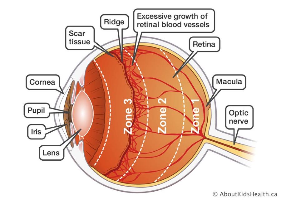
This condition manifests when irregular blood vessels develop in the retina, the light-sensitive layer of tissue located at the back of the eye. While certain infants with ROP experience mild cases that resolve without intervention, others may require treatment to safeguard their vision and prevent the onset of blindness.
Causes and Risk Factors of Retinopathy of Prematurity (ROP)
- ROP primarily occurs in infants born prematurely, especially those with a gestational age of less than 31 weeks.
- The blood vessels haven’t fully developed yet in the retina.
- Infants weighing less than 3 pounds at birth are at a higher risk of developing ROP.
- The administration of high levels of oxygen to premature infants, especially for an extended duration, is a significant risk factor for ROP development.
- Certain medical conditions and complications in premature infants, such as respiratory distress syndrome, can contribute to the development of ROP.
- Multiple blood transfusions in preterm infants may increase the risk of ROP.
- Infections during the neonatal period can be associated with an increased likelihood of developing ROP.
- There may be a genetic predisposition to ROP in some cases.
Understanding these causes and risk factors is crucial for identifying high-risk infants and implementing preventive measures, as well as for the early detection and management of ROP. Regular monitoring and appropriate medical care play key roles in mitigating the impact of these risk factors on the development of retinopathy of prematurity.

Signs and Symptoms of Retinopathy of Prematurity (ROP)
- Infants with ROP may exhibit unusual eye movements, such as rapid and irregular eye jerks.
- The presence of a white or cloudy appearance in the pupils, instead of the normal red-eye reflection, can be indicative of ROP.
- Excessive tearing or tearing that occurs when not associated with crying may be a symptom of ROP.
- Premature infants with ROP may display sensitivity to light, avoiding bright lights and reacting strongly to sudden changes in illumination.
- The impaired ability to visually track or follow objects with the eyes may suggest ROP.
- Strabismus, where the eyes do not align properly, can be a sign of ROP.
- Infants with ROP may show decreased responsiveness to visual stimuli or have difficulty fixing their gaze.
- Any sudden changes in the appearance of the eyes, including cloudiness or discoloration, should be promptly evaluated.
It’s essential for parents, caregivers, and healthcare providers to be vigilant for these signs and symptoms, especially in premature infants or those at risk for ROP. Regular eye examinations as part of neonatal care can aid in the timely identification of ROP-related changes.
Diagnosis
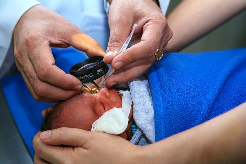
Diagnosing Retinopathy of Prematurity (ROP) involves specialized eye examinations conducted by an experienced ophthalmologist. These examinations, which typically begin shortly after birth, include the dilation of pupils using eye drops to enable a detailed examination of the retina.
The ophthalmologist uses specialized tools, such as a binocular indirect ophthalmoscope or a digital retinal camera, to capture images of the retina. The severity of ROP is then classified into stages based on the extent of abnormal blood vessel growth. Continuous monitoring through follow-up examinations is essential to track the progression of ROP and determine if intervention is necessary. Early diagnosis is critical for timely and effective management of the condition, ensuring the best possible outcomes for the infant’s vision.
Stages of ROP
The progression of Retinopathy of Prematurity (ROP) is categorized into five stages, allowing doctors to assess the severity of the condition and determine the appropriate course of action:
Stages 1 and 2 (Mild to Moderate ROP)
Babies in these stages often improve without treatment and can maintain healthy vision. Doctors closely monitor them to track the progression of ROP.
Stage 3 (Severe ROP)
Some infants with stage 3 ROP may spontaneously recover without intervention and go on to have healthy vision. However, others may require treatment to prevent abnormal blood vessels from damaging the retina and causing retinal detachment, a condition that can lead to vision loss.
Stage 4 (Partial Retinal Detachment)
Babies in stage 4 have partially detached retinas, necessitating treatment to address the severity of the condition.
Stage 5 (Complete Retinal Detachment)
In stage 5, the retina is completely detached. Even with treatment, infants in this stage may experience vision loss or blindness. Stages 4 and 5 are particularly serious, often requiring surgery, but vision loss may still occur despite treatment.
Retinopathy of Prematurity Treatment Options
Many cases of ROP resolve without treatment, but when intervention is necessary, several options exist to protect your child’s vision. Early treatment is crucial. Here are the common treatment options:
Laser Treatment
For advanced cases of ROP, laser treatment may be recommended. This procedure targets the sides of the retina to prevent the worsening of ROP and safeguard your child’s vision.
Injections of Anti-VEGF Drugs
Doctors can administer medications known as anti-VEGF drugs through injections into the baby’s eye. These drugs work by blocking the growth of abnormal blood vessels, a key factor in ROP.
Eye Surgery
For stages 4 or 5, where the retina is partially or completely detached, surgery becomes necessary. There are two main types:
1. Scleral Buckle Surgery
A flexible band is placed around the sclera (the white part of the eye). This band supports the detached retina until the eye grows normally. The band is later removed by the doctor.
2. Vitrectomy
Small openings are made in the eye wall to remove most of the vitreous (the gel-like fluid filling the eye), which is replaced with saline solution. The surgeon then removes scar tissue from the retina. Laser treatment may also be performed to treat and secure the retina in position.

How can vision loss be prevented?
The early prediction or control of premature birth poses significant challenges, but effective neonatal care coupled with timely screening and urgent laser therapy can substantially decrease the occurrence of blindness or visual impairment in infants.
Doctors and nurses can employ to mitigate the risk of retinopathy of prematurity (ROP) using the POINTS of Care approach: managing pain, judicious oxygen administration, infection prevention, promoting nutrition through breastfeeding, ensuring optimal temperature regulation, and implementing supportive practices like kangaroo care to enhance infant comfort and stability.
Early screening for ROP is imperative to identify infants at risk of developing severe, vision-threatening stages of the condition. Typically, screening is conducted by a skilled ophthalmologist within the neonatal unit using indirect ophthalmoscopy. Determining whom to screen and the timing of screening hinges on various factors, including the quality of neonatal care provided. In settings where care falls short, larger, more mature infants should also undergo screening, as they too can be susceptible to vision-threatening ROP.
Given that ROP manifests after birth, with onset occurring within the initial weeks of life, the initial screening examination should occur no later than 30 days post-birth. Subsequent follow-up screenings may be necessary, potentially even after the infant has been discharged from the neonatal unit. Each country must establish screening criteria tailored to its specific circumstances.
Prompt treatment is essential for all infants exhibiting sight-threatening stages of ROP and should be initiated within 48 to 72 hours.
Furthermore, diligent monitoring of all preterm infants is crucial, as they face heightened risks of other conditions that can result in vision loss. Such risks are amplified among infants with a history of ROP, particularly those who have undergone treatment. Refractive errors, including early-onset severe myopia, are the most prevalent conditions. Additionally, conditions such as strabismus and cerebral visual impairment occur at higher rates compared to full-term infants.
