To protect your vision from dangerous eye diseases like Glaucoma or macular degeneration, early and accurate diagnosis is the golden key. This is precisely where Optical Coherence Tomography (OCT) plays an indispensable role in modern ophthalmology.
OCT scanning (retinal optical coherence tomography) is a remarkable breakthrough, allowing doctors to “look deep” into the tiny layers of cells and nerve fibers in the eye without the need for surgery. This is a non-invasive, painless, and X-ray-free technique, ensuring absolute safety for the patient.
This article will help you understand what an OCT eye scan is, how it works, why it’s important, and who should undergo this technique. Let’s explore to proactively protect your precious eyes.
What Is OCT Eye Scan?
OCT eye scan (short for Optical Coherence Tomography) is a modern diagnostic imaging technique in ophthalmology. It helps doctors observe the detailed internal structures of the retina and optic nerve. This method operates based on the principle of light interference, allowing for high-resolution cross-sectional images of the retina, similar to a computed tomography (CT scan) but completely painless and non-invasive.
OCT is particularly important in early detection and monitoring of eye conditions such as: macular degeneration, macular edema, glaucoma (increased intraocular pressure), retinal detachment, maculitis, and eye complications due to diabetes. It is an essential tool for maintaining visual health.
>> Modern Eye Examination and Diagnostic Methods
How Retinal Optical Coherence Tomography (OCT) Works
The operating principle of OCT scanning is based on the use of safe infrared light waves.
Light “Scans” Detailed Eye Structures
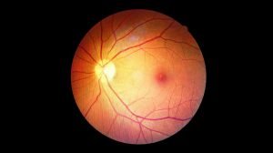
The OCT device emits a beam of infrared light that shines into the eye. When this light encounters different tissue layers within the eye (such as the retina, nerve fibers, blood vessels), it reflects back to the machine. The OCT machine measures the time and intensity of the reflected light to construct extremely detailed 2D or 3D cross-sectional images, with a resolution measured in micrometers (µm) – smaller than a human hair.
Near-infrared light does not cause glare or discomfort. This technology is particularly effective in imaging sensitive structures like the retina and optic nerve head, where even the smallest changes can be signs of serious pathology.
The Importance of OCT in Diagnosing Eye Diseases
OCT scanning is a silent “hero,” helping to detect many dangerous eye diseases early that can lead to blindness.
Early Detection of Glaucoma (Increased Intraocular Pressure)
Glaucoma is often called the “silent thief of sight” because it usually has no symptoms in its early stages. OCT scanning plays a crucial role in early glaucoma detection by measuring the thickness of the retinal nerve fiber layer (RNFL) and assessing the optic nerve head. Even when vision is not yet affected, OCT can identify subtle thinning of the nerve fibers – the earliest sign of glaucoma.
- Quantitative assessment: OCT provides metrics that allow doctors to compare with normative data, leading to an accurate diagnosis of glaucoma status.
- Monitoring progression: The ability to compare serial OCT scans helps track changes over time, evaluate treatment effectiveness, and adjust treatment plans promptly.
Diagnosis and Monitoring of Retinal Diseases
The retina is the most sensitive neural tissue at the back of the eye. OCT scanning is an indispensable tool in the diagnosis and monitoring of retinal diseases, which are leading causes of vision loss.
- Age-related macular degeneration (AMD): OCT clearly visualizes the macular layers, enabling early detection of dry AMD (drusen) and is particularly important in diagnosing wet AMD (characterized by neovascularization, hemorrhage, and edema).
- Diabetic Retinopathy: OCT measures macular thickness, detects diabetic macular edema (DME), and helps in planning effective treatment strategies.
- Macular hole and epiretinal membrane: OCT clearly visualizes these structures, assessing the degree of traction to determine appropriate surgical intervention.
>> Regular Eye Exams for Diabetics: Your Strongest Shield Against Dangerous Vision Loss
Applications of OCT in Other Eye Conditions
Beyond the retina and glaucoma, OCT scanning has several other important applications:
- Cornea and anterior segment: OCT performs cross-sectional imaging of the cornea, evaluates corneal pathologies, or assists in diagnosing angle-closure glaucoma using anterior segment OCT (AS-OCT).
- Optic nerve: Aids in diagnosing non-glaucomatous optic neuropathies.
- Pre- and post-operative screening: Assesses eye structures before crucial surgeries like cataract surgery, LASIK, and monitors post-operative outcomes to ensure safety and effectiveness.
Who Should Get an OCT Eye Scan?
OCT eye scans are recommended for many individuals, especially those with high risk factors or existing eye symptoms.
You should consider getting an OCT eye scan if:
- You are over 40 years old: Especially if you have a family history of Glaucoma or diabetes.
- You have diabetes: To monitor and enable early detection of diabetic retinopathy complications.
- You have a family history of Glaucoma: Regular screening helps in early detection and preventive measures.
- You have Glaucoma or suspected Glaucoma: For accurate diagnosis, assessment of damage extent, and monitoring treatment efficacy.
- You have symptoms suggestive of retinal disease: Such as blurred vision, seeing black spots, or distorted images.
- You are undergoing treatment for retinal conditions: To evaluate the effectiveness of medications or other treatment methods.
- You need assessment of the cornea or anterior chamber angle.
- You are having a general eye health check-up at a certain age.
The benefits of OCT scanning include early disease detection, accurate diagnosis, effective treatment monitoring, and a safe, quick procedure, providing peace of mind to patients.
OCT Eye Scan Procedure
The OCT scan procedure is very quick, usually taking only about 5-10 minutes for both eyes.
Preparing for the Scan
- You do not need to fast.
- Inform your doctor about your medical history and any medications you are currently taking.
- Remove contact lenses before the scan.
- Pupil dilation may be required: If dilating eye drops are administered, your vision will be temporarily blurred and you will be sensitive to light. Therefore, you should wear sunglasses and avoid driving after the scan.
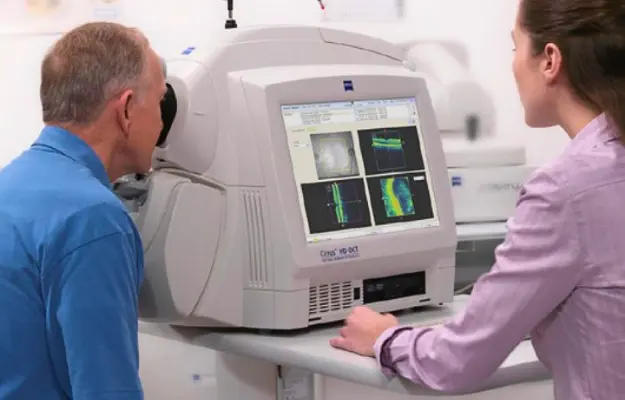
During the Scan
You will sit in front of the machine, place your chin on a chin rest, and look at a fixed point. The machine will emit a beam of infrared light that scans across your eye. You just need to keep your eye still for a few seconds. The entire process is painless and comfortable.
After the OCT Scan
After the scan, the ophthalmologist at European Eye Center will analyze the images and graphs, explain the results, and provide a diagnosis. Based on this, the doctor will advise on a treatment plan or schedule for follow-up visits, helping you proactively manage your eye health.
Choosing a Reputable OCT Scan Facility
To ensure the effectiveness and safety of your OCT scan, choose a reputable medical facility with:
- Modern OCT equipment: Ensuring sharp, accurate images for the best diagnostic support.
- Experienced doctors and technicians: Professional interpretation and analysis of results, providing precise advice.
- Comprehensive examination process: Combining OCT with other eye examination steps to get a holistic view of your eye health.
- Standardized facilities and professional services: Creating comfort and trust for patients.
Investing in a quality eye care facility is an investment in your long-term eye health.
Conclusion
Optical Coherence Tomography (OCT) is an indispensable “gold standard” in the diagnosis and monitoring of eye diseases. Its ability to visualize even the earliest microscopic lesions has helped OCT scanning save the vision of millions worldwide.
Proactively protect your eyes by understanding OCT scanning and seeking reputable ophthalmology specialists for consultation and examination when needed. Early detection is always the key to effective treatment and maintaining clear, healthy vision for a long time.