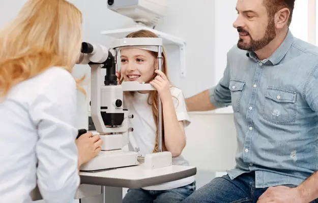
In an increasingly digital world, proper eye care has become more crucial than ever. Our eyes are constantly working, exposed to blue light from electronic screens, and facing a myriad of other potential risks. To protect and maintain vision, regular eye examinations using modern eye examination and diagnostic methods are absolutely essential. The groundbreaking advancements in ophthalmology have introduced a host of sophisticated technologies that not only help doctors accurately determine refractive errors but also enable the early detection of dangerous eye diseases, and even systemic conditions like diabetes, hypertension, or autoimmune diseases that manifest in the eyes.
This article serves as a comprehensive guide, synthesizing and introducing the most common modern eye examination and diagnostic methods available today. We will delve into the operating principles, clinical applications, and significance of each technique, from fundamental screening steps to specialized diagnostic imaging technologies. This will provide you with a holistic understanding and help you better prepare for your next eye exam.
Methods for Assessing Visual Acuity and Refractive Errors
These are the fundamental and indispensable steps in every eye examination. They help determine your visual ability and identify common refractive errors such as myopia (nearsightedness), hyperopia (farsightedness), and astigmatism.
Visual Acuity Test
This is the most basic method for measuring your eye’s ability to see detail. A doctor will ask you to read letters or symbols on a chart from a specific distance.
- Snellen Chart: This is the most common type of chart, featuring rows of letters that decrease in size. The result is typically expressed as a fraction (e.g., 20/20, 20/40), where 20/20 represents normal vision, meaning you can see clearly at 20 feet what a person with normal vision can also see at 20 feet.
- ETDRS Chart: This is considered the gold standard chart for clinical studies, providing a more accurate and reliable assessment of visual acuity, especially in individuals with low vision.
- Advantages: It’s simple, quick, and serves as an initial screening step to give an overall assessment of the patient’s vision.
>> For more information on advanced solutions that support accurate vision testing and optimize the eye examination process, you can visit Blincq Solutions.
Refraction Test
This method is used to accurately determine your eye’s refractive error, which is essential for prescribing the correct glasses or contact lenses.
- Auto-refractor: This device uses infrared light to automatically measure the eye’s refractive parameters. It quickly and accurately provides an initial reading that serves as a baseline for the ophthalmologist.
- Subjective Refraction: After the initial reading, the doctor uses a device called a phoropter to fine-tune the prescription. During this process, the doctor will ask for your feedback on the clarity of the image as different lenses are presented, aiming to find the best possible prescription for both eyes.
- Cycloplegic Refraction: For children and young adults, the eye’s accommodative (focusing) muscles are very strong and can lead to inaccurate readings. Therefore, a doctor may use cycloplegic eye drops to temporarily relax these muscles, allowing for a measurement of the true refractive error.
>> Modern Treatments and Surgical Methods for Eye Conditions
Advanced Diagnostic Imaging Methods
Thanks to technological advancements, diagnostic imaging has revolutionized ophthalmology, enabling doctors to visualize detailed internal eye structures that were previously inaccessible.
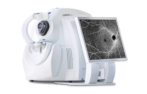
Optical Coherence Tomography (OCT)
OCT is one of the most powerful and widely used diagnostic imaging technologies today. The OCT machine uses light waves to create high-resolution cross-sectional images of internal eye structures, particularly the retina, optic nerve, and macula.
- Operating Principle: OCT is similar to an ultrasound, but instead of sound waves, it uses light waves to create images. A beam of light is directed into the eye, and the reflected light waves are captured. Based on the time and intensity of the reflections, a computer reconstructs a detailed, non-invasive, and painless cross-sectional image. The resolution of OCT can be as fine as 10-20 microns, allowing doctors to study eye structures at a microscopic level.
- Clinical Applications:
- Early Diagnosis of Retinal Diseases: Diabetic retinopathy, macular degeneration, macular holes, and epiretinal membranes. OCT helps assess the degree of swelling, hemorrhage, and other damage.
- Assessment of Optic Nerve Damage: This is crucial for diagnosing and monitoring glaucoma. OCT quantitatively measures the thickness of the retinal nerve fiber layer.
- Pre- and Post-operative Examination: It helps evaluate eye structures before surgery and monitor the recovery process afterward.
Fluorescein Angiography (FA)
FA is a dynamic imaging technique that allows doctors to visualize the intricate vascular system of the retina and choroid.
- Operating Principle: The doctor injects a fluorescent dye into a vein in your arm. A specialized camera then takes a rapid series of photographs of the retina as the dye circulates through the blood vessels. The doctor can directly observe areas of leakage, blockage, or abnormal blood vessels.
- Clinical Applications:
- Detection and Evaluation of Vascular Abnormalities: Retinal artery/vein occlusion, vascular leakage (in diabetic retinopathy).
- Diagnosis of wet macular degeneration, helping doctors pinpoint the location and size of abnormal blood vessels to guide treatment.
- Diagnosis of Central Serous Chorioretinopathy.
Ocular Ultrasound
Ocular ultrasound uses high-frequency sound waves to create images of the internal structures of the eye. It is especially useful when other methods are ineffective due to an opaque internal environment (e.g., dense cataracts, vitreous hemorrhage).
- Operating Principle: An ultrasound probe is placed on the eyelid with a special gel or directly on the eyeball after a local anesthetic is applied. The probe emits sound waves, which reflect off the internal structures of the eye. The computer captures these reflected waves to create a detailed image.
- Clinical Applications:
- Detection of retinal detachment, vitreous hemorrhage, or tumors in the back of the eye.
- Assessment of the lens and vitreous humor.
- Localization of foreign bodies after an injury.
- Measurement of the eyeball’s length (A-scan ultrasound) to calculate the power of an intraocular lens before cataract surgery.
Other Essential Diagnostic Methods
In addition to the imaging techniques above, several other methods provide a comprehensive picture of your eye health.
Tonometry
Tonometry measures the intraocular pressure (IOP) within the eye. This measurement is a critical step in diagnosing and monitoring glaucoma. High IOP is the main risk factor for optic nerve damage.
- Common Methods:
- Non-contact tonometry: Uses a gentle puff of air to the cornea. This method is quick, painless, and does not require anesthetic drops.
- Applanation tonometry: Uses a small probe that gently touches the cornea after it has been numbed with drops. This method provides more accurate results and is considered the gold standard.
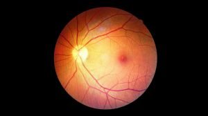
Fundus Examination
This is a classic and essential technique that allows the doctor to directly observe the retina, optic disc, and blood vessels at the back of the eye.
- Procedure: The doctor uses a device with a light and a magnifying lens to examine these structures. This technique is often performed after dilating the pupil with eye drops for a clearer view.
- Advantages: Allows for direct observation and detection of signs of retinal diseases, glaucoma, hypertensive retinopathy, and many other conditions.
Visual Field Test
The visual field is the entire area of space that the eye can see without moving the head. A visual field test helps detect blind spots or areas of peripheral vision loss.
- Application: This is a crucial test for diagnosing and monitoring glaucoma, as the disease often causes a gradual, progressive loss of peripheral vision.
Conclusion
Modern eye examination and diagnostic methods not only help accurately determine refractive errors but also serve as a vital shield, protecting the eyes from dangerous diseases. From simple visual acuity tests to advanced imaging technologies like OCT and FA, each method plays a critical role in providing a comprehensive assessment of eye health.
Regular eye exams, especially for high-risk individuals (the elderly, people with diabetes, severe myopia, or a family history of eye disease), are the most proactive and intelligent step you can take to preserve your vision. Entrust your care to expert ophthalmologists and modern technology to ensure your eyes remain healthy and clear.