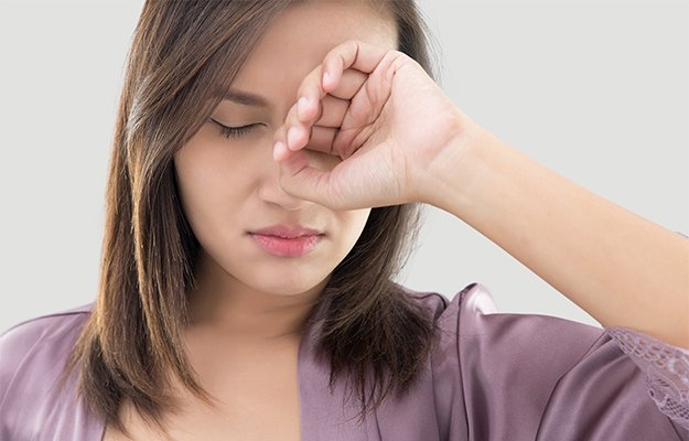
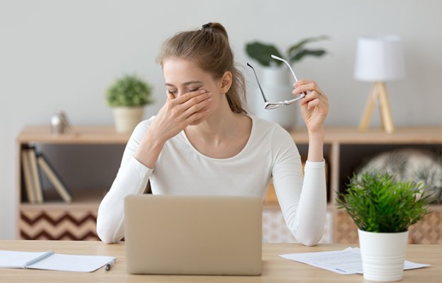
Have you ever noticed your eyelid starts to quiver uncontrollably for no apparent reason? Many of us experience eye twitching during our lifetime for a variety of reasons. Learn about eye twitching and some at-home tips to stop eye twitching.

What Is Eye Twitching?

Eye twitching, also called myokymia, is a painless condition that occurs when your eyelid muscles contract involuntarily. It only affects a limited amount of fibers from the eyelid muscles and it can range from barely noticeable to bothersome.

Female-green-eye-close-up-How-to-Stop-Eye-Twitching-ss-feat

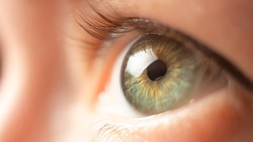
So what does eye twitching feel like? Most people describe it as a slight sensation of tug on the lower or upper eyelid, usually unilaterally (one eye only).

Eye twitching is very common and usually resolves on its own, within a few seconds to a few minutes, but may be recurrent over a few hours or days. In some cases, a twitch can last longer than expected, but it is not a reason for concern.

Although eyelid twitching is rarely a sign of a severe condition, it can turn into a nuisance when episodes of twitching happen more frequently than usual.

Besides eye twitching, there are 2 other conditions that manifest with involuntary eye lid movement:

  • Benign essential blepharospasm: increased blinking of both eyes which can progress to eyelids being squeezed shut
  • Hemifacial spasm: involves twitches of more muscles on one side of the face, including the eyelid

Why Is My Eyelid Twitching?

No one knows the exact cause behind eye twitching. Health experts believe that several factors play a role in spontaneous eyelid muscle contractions.  Aside from general stress and fatigue,  possible causes of eye twitching are:

  • Environmental irritants (pollutants, dirt, bright light, wind)
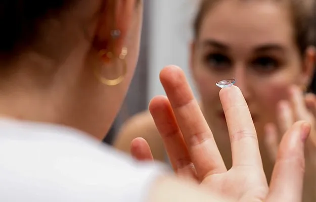
  • Eye strain or irritation
  • Caffeine intake
  • Excessive drinking and smoking
  • Dry eyes
  • Medication side effects
  • Uveitis
  • Blepharitis
  • Migraine
  • Light sensitivity

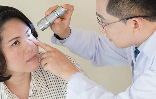
When to see a doctor

Usually eye twitches resolve on their own within a few hours to a few days with rest, stress relief and less caffeine intake.

If the twitches are persistent for a few weeks or are accompanied by other symptoms, you should check in with your doctor. When accompanied by other symptoms, twitching might be a sign of a brain or nervous system disorder, like Bell’s palsy, cervical dystonia, multiple sclerosis and Parkinson’s disease, to name a few.

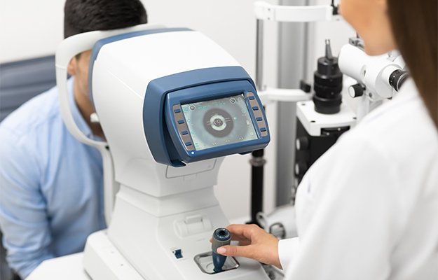
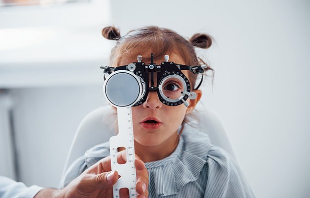
Visit an eye doctor as soon as possible if your eyes keep twitching for a few weeks or when one of the following symptoms appear:

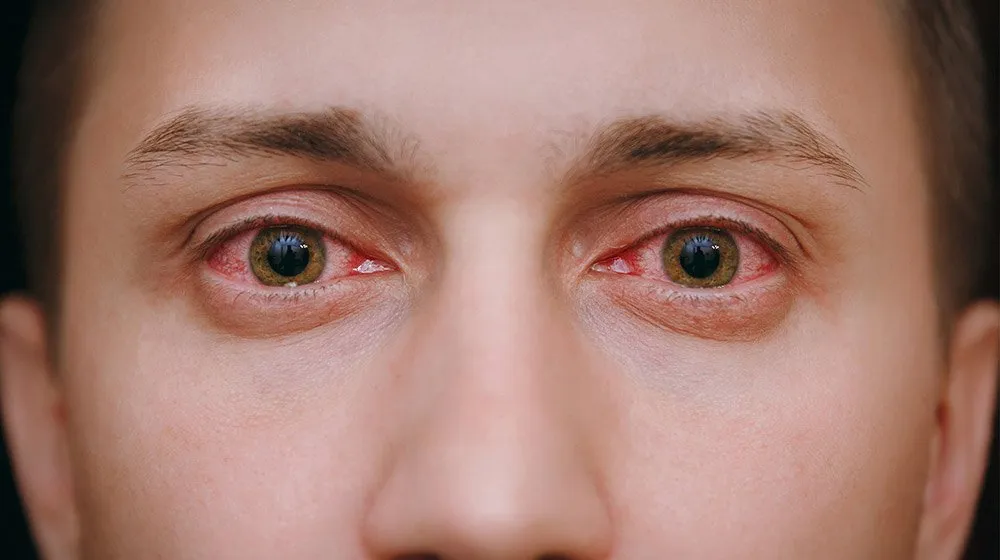
  • Your eye is red with abnormal discharge
  • Drooping eyelids
  • Twitching in other parts of your face or body
  • Your eyelid closes completely with each twitch

7 Tips to Stop Eye Twitching

Although eye twitching resolves on its own, there are some home remedies you can try to relieve eye twitching:

Try Blinking Hard

woman-meditates-takes-break-after-hard-workTry-Hard-Blinking-ss-body - How to Stop Eye Twitching

Close your eyes as tight as possible. This will help spread tears evenly over the eye surface, preventing your eyes from drying out and relieving twitching.

Let Your Eyes Rest

top-view-of-bearded-man-sleeping-on-bed-Let-Your-Eyes-Rest-ss-body

People under a lot of stress or very fatigued, are prone to eye twitching. Taking a nap or de-stress can help restore your eyes more quickly.

Use a Warm Compress

Using a warm compress or warming eyepatch and applying it to the eyes can relax the muscles surrounding them.

Limit Caffeine Intake

Caffeine is a stimulant that recharges your body. Too much caffeine, however, either in your coffee, tea, or energy drink, can cause twitches to appear.

Limit your caffeine intake gradually and keep hydrated with water.  Dehydration is also linked with eye twitching.
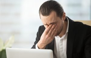
Cut Down on Stress

High-stress levels have a negative impact on your overall health and are also the major cause of eyelid twitches. Try meditation or breathing exercises, and reorganize your work schedule to limit stress.

Moisturize Your Eyes

There is a link between dry eye syndrome and eye twitches. Hydrating eye drops or artificial tears are a good idea to refresh and moisten your eyes.

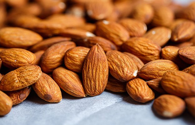
Consider Supplements

Proper muscle function depends on different vitamins and minerals. An imbalance in these nutrients can be a cause of eye twitching. Especially vitamin B12, vitamin D and magnesium levels are important.

Consult with your eye doctor first before taking any supplements.

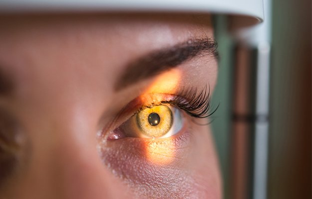
If you are experiencing constant eye twitching accompanied by an uncomfortable feeling, schedule an exam with us today to find the best treatment for your eyes.

Every patient deserves top-quality eye care from us. European Eye Center focuses on providing Western-standard services and determining appropriate treatment plans to help patients restore their vision in a feel-like-home atmosphere.

We are happy to help if you have questions about how to stop eye twitching. Don’t hesitate to connect with us at appointment@europeaneyecenter.com.

Contact us today to schedule an eye care service!

Eye strain, UV exposure, and genetics play a role in eye health and vision but studies show that vitamins and nutrients can promote eye health and prevent eye conditions like age-related macular degeneration. Nutrients and vitamins like lutein, zeaxanthin, omega-3 fatty acids, vitamins C and E, and zinc are essential in maintaining good vision and healthy eyes.

Which Foods Are the Best for Eye Health?

Fish

snapper-fish-steamed-with-soy-sauce-Fish-ss-body

Fish and seafood are the best natural sources of omega-3 fatty acids or healthy fats. Omega-3 fatty acids are essential for retina health and vision development. They can also help prevent dry eye syndrome.

Adding fish to your diet, especially salmon, trout, mackerel, tuna, and sardines, will help maintain eye health.

Carrots

boiled-carrots-Carrots-ss-body

For decades, carrots have been known to be beneficial for eye health. Carrots contain beta-carotene, a substance that the body converts into Vitamin A, an important nutrient for eye health. An extreme lack of vitamin A can cause blindness. Vitamin A can also help prevent the formation of cataracts and macular degeneration. Besides helping fight infections, vitamin A is also essential in preventing dry eye, corneal scarring, and night blindness.

Carrots are easy-to-eat food that can be a snack or turned into soup, salad, and cakes. Do remember that the beta-carotene in carrots might cause your skin to become discolored if consuming too many. So aim for no more than 1 or 2 servings every day.

Eggs

Fluffy-omelette-from-whipped-eggs-with-cheddar-cheese-Eggs-ss-body

Egg yolks are rich in zinc, lutein, zeaxanthin, and vitamin A,  all essential for maintaining eye health.

Vitamin A protects the cornea, while zinc supports the retina’s health and improves night vision. Zeaxanthin and lutein reduce the risk of developing age-related macular degeneration and cataracts, as well as other chronic eye diseases.

Nuts and Seeds

roasted-sweet-almonds-lie-in-a-large-heap-Almonds-ss-body

Nuts and seeds, particularly almonds, contain omega-3s and vitamin E, which are beneficial for healthy eyes.

Vitamin E, which protects against unstable molecules, is found in almonds. These molecules attack and harm healthy tissue. Regular vitamin E intake can also help prevent cataracts and age-related macular degeneration.

Dark Green Leafy Vegetables

green-vegetables-on-wooden-surface-Dark-Green-Leafy-Vegetables-ss-body

Kale and spinach are superfoods because they contain essential vitamins, nutrients, and minerals. They also support eye health.

Dark green, leafy vegetables, such as kale, spinach, and broccoli, contain a lot of antioxidants like lutein and zeaxanthin, which help prevent severe eye conditions. These two antioxidants are found in high amounts in the macula and retina, and by serving as a light filter, lutein and zeaxanthin shield the retina from sunlight damage.

Other Tips to Boost Your Eye Health

  • Use sun protection, such as sunglasses or hats, to protect your eyes.
  • Limit screen time and blue light from the television, phone, or computer.
  • Stop smoking because the smoke is linked to vision loss, cataracts, and macular degeneration.
  • Get regular eye exams to evaluate your eye health and detect any possible eye problems. Visiting an ophthalmologist frequently is an excellent preventive health habit you should consider.

If you or your family members suffer from any eye conditions, schedule an exam today to find the best treatment for their eyes.

Every patient deserves top-quality eye care from us. European Eye Center focuses on providing Western-standard services and determining appropriate treatment plans to help patients restore their vision in a feel-like-home atmosphere.

We are happy to help if you have questions about foods for eye health. Don’t hesitate to connect with us at info@europeaneyecenter.com.

Contact us today to schedule an eye care service!

Diabetes is a metabolic disorder that causes too high blood glucose levels in millions of people worldwide.The prevalence is rising dramatically in the last decade, especially in low- and middle-income countries.

ophthalmologist-checking-eye-vision-Visit-Eye-Doctor-for-Annual-Eye-Exam-ss-body

Diabetes can give rise to various health complications all over your body since it affects all the small blood vessels.

Diabetic retinopathy is a common eye complication of diabetes. However, did you know you can reduce the risk of developing diabetic retinopathy with discipline and simple daily practices?

Here’s what you can do to reduce the risk of developing diabetic eye disease:

Visit your Eye Doctor for an Annual Eye Exam

Everyone should have regular eye exams, but for diabetes patients, it’s especially important to have annual eye exams. Early stages of diabetic retinopathy can be asymptomatic for a long time, but that doesn’t mean no damage is being done. When you do experience symptoms like blurry vision, flashes, dark spots, or vision loss, contact your ophthalmologist right away.

An annual eye exam is important to keep track of the disease progression and to intervene on time when necessary.

Furthermore, diabetes affects the small blood vessels in your entire body, not just in your eyes. The difference is, that the blood vessels in your eyes can be seen directly by your eye doctor.

If changes due to diabetes appear in the eyes, similar changes will also be present in areas like your kidneys, feet, and all other parts of the body with blood vessels. Timely diagnosis leads to more successful treatments and better outcomes, whatever the part of the body that is affected.

Monitor Your Blood Sugar

Continuous high blood sugar levels are bad for the small blood vessels in your body, especially in your eyes. Managing your blood glucose levels is essential in reducing the risk of diabetic retinopathy and the associated vision loss.

A glucose meter is one of the methods to take control of your blood sugar levels at home.

More specialized blood tests like HbA1C reflect your blood glucose levels over a longer period of time.

With glucose monitoring, you can adjust your medication accordingly and keep levels under control.

Stick to a Healthy Diet

A healthy diet is another significant way to help manage blood sugar levels.

The most harmful foods for diabetes are sugary and sweet foods, including fast foods, soda, cereals, dried fruits, packaged nuts, juices, flavored yogurts, but also white bread, pasta and rice.

So what foods are safe to eat for people with diabetes? Here are some of the best diabetic-friendly foods:

  • Fatty fish
  • Dark green vegetables
  • Eggs
  • Beans
  • Whole grains
  • Plant-based proteins
  • Greek yogurt
  • Nuts
  • Flaxseeds
  • Garlic
  • Chia seeds
  • Avocados

Manage Your Cholesterol Levels

If you have diabetes, you will likely have lower levels of HDL cholesterol (good cholesterol) and higher levels of LDL cholesterol (bad cholesterol). High blood glucose levels damage the wall of the small blood vessels and make it more likely for cholesterol to stick to them, increasing the risk of narrowing them or making them blocked. Check and control your cholesterol levels regularly with diet and medication, if needed.

Exercise

Exercise is very beneficial for controlling blood sugar levels. It increases your sensitivity to insulin and lowers blood sugar levels. Physical exercise also reduces your cholesterol, lowers your blood pressure and your risk of heart disease.

You don’t have to do intense workouts. Simple, light exercises, like walking, jogging, swimming, and cycling, can have positive effects. It’s always best to consult your doctor before starting any new exercise regimen.

Quit Smoking and Alcohol

The negative health effects of smoking are well documented and for diabetes patients, smoking makes managing diabetes also more difficult. Nicotine increases blood sugar levels and often more insulin is needed to lower these. Smoking also increases the risk of developing type 2 diabetes by 30-40%.

Diabetes patients can drink 1-2 glasses of wine, but need to be aware of the alcohol’s effect on the body. Moderate amounts of alcohol usually cause a rise in blood glucose levels, but larger amounts can actually decrease the levels, sometimes to dangerously low levels. Excess alcohol intake is associated with an increased risk of type 2 diabetes.

Wear Eye Protection

UV exposure is linked to cataracts and age-related macular degeneration. Wear protective eyewear when going outside and wear sunglasses that block 100% of damaging UV rays.

Take Breaks When Using a Computer

man-relaxing-on-chair-sitting-at-table-Take-Breaks-When-Using-a-Computer-ss-body

Long hours of using a computer or other digital device cause digital eye strain.

The 20-20-20 rule is a simple practice that you can follow to proactively protect your eyes from digital eye strain and other eye problems: every 20 minutes, take a 20-second break by looking at an object at least 20 feet away.

De-stress

Too much stress and anxiety are linked to higher risks of retinopathy.

Stress causes the release of cortisol, the stress hormone, which is linked to inflammation and leakage of small blood vessels, exacerbating the damage caused by diabetes. Stress can also cause or increase insulin resistance and higher blood glucose levels. All these factors increase the risk of or exacerbate diabetic retinopathy.

Seek Help When Needed

Keeping up with your vision allows you to notice changes in your vision early. Dark spots in your vision field can indicate leaking blood vessels in the retina or an increase in flashes or floaters may indicate a retinal detachment. Always contact your ophthalmologist when symptoms arise. This practice allows your doctor to devise an appropriate treatment plan to protect and preserve your vision when you have diabetes.

If you have diabetes and want to take control of your eye health throughout the years, schedule a visit to the European Eye Center today.

Every patient deserves top-quality eye care from us. European Eye Center focuses on providing Western-standard services and determining appropriate treatment plans to help patients restore their vision in a feel-like-home atmosphere.

We are happy to help if you have questions about diabetic retinopathy. Don’t hesitate to connect with us at info@europeaneyecenter.com.

Contact us today to schedule an eye care service!

In this article, we will learn more about diabetic retinopathy – a common complication among diabetic people – including causes, symptoms, diagnosis and treatments.

A man having eye pain, wearing a suit_SS

What Is Diabetic Retinopathy?

Diabetic retinopathy is an eye condition that develops as a complication in people with diabetes.

The condition is known to progressively damage the blood vessels of the retina. The walls begin to weaken and the blood vessels start leaking blood and fluid into the retina.

Without prompt intervention, diabetic retinopathy can cause permanent vision loss even though it has little to no symptoms in the early stages.

Causes

At first glance, most people think that diabetes and eye health are not related. However, high or fluctuating blood glucose levels caused by diabetes can obstruct the blood supply to the retina, because of damage to the small blood vessels.

Obstructed blood supply results in oxygen deprivation of the retina, which in turn triggers the formation of new, abnormal blood vessels. These new blood vessels are of bad quality and leak easily.

Both type 1 and 2 type diabetes, as well as gestational diabetes (diabetes during pregnancy), have a risk of developing DRP. The risk increases as a result of:

  • Prolonged diabetes
  • Poor management of blood sugar level
  • High cholesterol
  • High blood pressure (hypertension)
  • Unhealthy lifestyle (smoking, substance use, drinking)
  • Pregnancy
  • Being Native American, Black, or Hispanic

Stages of Diabetic Retinopathy

Diabetic retinopathy is classified into two main stages: non-proliferative and proliferative diabetic retinopathy.

Non-Proliferative Diabetic Retinopathy (NPDR)

Non-proliferative diabetic retinopathy is the early stage of diabetic retinopathy.

Most people with diabetes type 1 and 60% of people with type 2 have NPDR but are usually unaware because the symptoms are relatively mild.

With NPDR,  the blood vessels become tortuous and can start to leak, the retina swells up due to excess fluid. Sometimes the fluid builds up in the central area of the retina causing macular edema. This is the leading cause of blurred vision in people with diabetes.

Proliferative Diabetic Retinopathy (PDR)

Proliferative diabetic retinopathy is an advanced and more severe stage of diabetes-related eye disease. It occurs especially if no action is taken to manage NPDR.  The blood vessels close off, cutting off the blood supply to the retina. This triggers the growth of new, abnormal blood vessels which leak blood and fluid into the vitreous, leading to eye floaters or vitreous hemorrhage.

The formation of scar tissue can lead to retina detachment or glaucoma.

Symptoms

women-in-purple-robes-have-itching-in-the-eye-Symptoms-ss-body

As mentioned above, not many people with diabetes realize they have diabetic retinopathy.

However, when it worsens, they may notice the following symptoms:

  • Blurry vision
  • More presence of floaters
  • Fluctuating vision – from blurry to clear and vice versa
  • Have problems seeing things at night
  • Dark areas in your vision
  • Vision loss

Diagnosis

To diagnose DRP your eye doctor will, besides taking a medical history, perform a comprehensive eye exam using drops to dilate the pupil and look at the retina.

Optical coherence tomography (OCT), a non-invasive imaging test, allows one to visualize cross-sections of the retina.

Treatments

Different treatments are available for diabetic retinopathy depending on specific problems and include:

  • Medicines: Anti-VEGF injections into the eye stop the growth of new blood vessels.
  • Laser treatment: This minimally invasive treatment seals or destroys leaking blood vessels in the retina.
  • Vitrectomy: Vitrectomy is a surgical procedure used for complications of proliferative diabetic retinopathy like retina detachment, non-clearing vitreous hemorrhage or macular traction.

Diabetes does not have to lead to vision loss. Diabetes management and yearly eye exams by a specialized ophthalmologist are the most important tools for preventing complications of diabetic retinopathy and preserving vision.

If you have diabetes and want to take control of your eye health, get an eye exam today at EEC.

Every patient deserves top-quality eye care from us. European Eye Center focuses on providing Western-standard services and determining appropriate treatment plans to help patients restore their vision in a feel-like-home atmosphere.

We are happy to help if you have questions about diabetic retinopathy. Don’t hesitate to connect with us at info@europeaneyecenter.com.

Contact us today to schedule an eye care service!

What is Amblyopia?

preteen-handsome-boy-play-squinting-trick-with-his-eyes-and-fingers-close---Diagnoses-and-Treatments---ss-body

Amblyopia (also called lazy eye) is poor vision in one eye. It develops in early childhood when the brain receives signals from both eyes that are too different.

The brain becomes confused and can not fully recognize the sight of the weaker eye. The brain prefers the image from the stronger eye and, over time, will only use the sight from that eye, while the vision of the weaker eye worsens.

It’s called lazy eye because the sight from the weaker eye is not being used by the brain, making the weaker eye even weaker over time.

Amblyopia starts to develop in early childhood, and it’s estimated that 3 to 6 out of 100 children have it. If left untreated, it can lead to permanent vision loss. Luckily early treatment works well.

What are the Symptoms of Amblyopia?

Unless your child has strabismus (cross-eyed) or a wandering eye, it is not obvious they have amblyopia. Your child might also not be aware that one eye is weaker than the other.

Signs that your child may have amblyopia are:

  • Poor depth perception (your child may appear clumsy)
  • Squinting
  • Closing 1 eye
  • Tilting his head

Most of the time, parents are not aware their child has amblyopia. Early treatment is crucial. That’s why kids need at least one thorough vision screening between the ages of 3 and 5.

What Causes Amblyopia?

Amblyopia occurs when the signals from the eyes to the brain are not similar. The brain will favor the best signal and start to ignore the signal from the weaker eye.

This can happen in the following cases:

  • A significantly higher refractive error in one eye (vision in one eye is a lot weaker than the other eye’s vision)
  • Strabismus or cross-eyedness. In this case, if one eye points in the wrong direction, it might drift in, out, up, or down.
  • Childhood Cataracts. Babies can be born with cataracts or can develop cataracts later. The cloudiness of the lens blurs the vision.
  • Ptosis or droopy eyelid. This can partially or entirely block the vision of one eye.

How is Amblyopia Treated?

The treatment of amblyopia starts with correcting the vision problem that’s causing the amblyopia.

So when a child is near or farsighted or has astigmatism, the eye doctor will prescribe eyeglasses or contacts. When cataracts cause a vision problem, surgery must be performed to remove cataracts.

Correcting the vision problem is not enough. When the vision problem is corrected, the brain has to be retrained in using the weaker eye. That way, the weaker eye will become stronger.

This can be achieved by wearing an eyepatch on the stronger eye for a few hours daily. The other option is to use Atropine drops once daily in the stronger eye. The Atropine drops cause temporary blurry vision and force the brain to use the weaker eye. Another option is to wear glasses with a lens that blurs the vision in the stronger eye.

This retraining of the brain usually takes a couple of months. After the vision in the weaker eye has strengthened, it will still be necessary to continue the eye patching temporarily during the next few years. Otherwise, the eye might weaken again.

Early treatment is essential and most effective when started before age 7. If left untreated, amblyopia may lead to permanent vision loss, so schedule your child’s first thorough vision screening between the ages of 3 and 5.

Every patient deserves top-quality eye care from us. European Eye Center focuses on providing Western-standard services and determining appropriate treatment plans to help patients restore their vision in a feel-like-home atmosphere.

We are happy to help if you have questions about amblyopia (lazy eye). Don’t hesitate to connect with us at appointment@europeaneyecenter.com.

Contact us today to schedule an eye care service!

You always follow the contact lenses cleaning guidelines and wear contacts without issues. But suddenly, symptoms such as blurry vision, grittiness, redness, pain, tearing, discharge, or light sensitivity, occur. You developed an eye infection.

Close-up-of-two-annoyed-red-blood-eyes-of-male-Contact-Lenses-and-Eye-Infections-ss-feat

Why, despite following cleaning guidelines, did you develop an infection?

What is the risk of a contact lens-related eye infection?

What are the symptoms of contact lens-related eye infections?

What should you do when developing an infection after wearing contact lenses?

Check this post for the solution.

What Causes Eye Infections From Contact Lenses?

Despite correct contact lens cleaning routines, developing an eye infection is always possible. Some of the causes are:

  • Bacteria or viruses (having a cold, for example, can lead to adeno virus eye infection)
  • Sleeping in your contact lenses
  • Using extended-wear lenses
  • Reusing contact lens solution or not keeping contact lens cases clean

Cleaning your contact lenses correctly is necessary to keep your eyes healthy. However, if not done properly, it will create an environment for growing germs like bacteria, viruses, fungi, or parasites on the lenses, and these germs can cause eye infections.

What Are The Risks Of Contact Lens-Related Eye Infections?

Because the contact lens sits on your cornea, the most common contact lens-related eye infection is keratitis, or infection of the cornea.

When severe enough, the cornea can develop a scar, impairing your vision, and sometimes a cornea transplant is necessary.

What Are The Symptoms Of Contact Lens-Related Eye Infections?

a-man-suffers-from-allergies-and-wipes-his-watery-eyes-Causes-of-Contact-Lens-Related-Infections-ss-body

  • Blurry vision
  • Grittiness
  • Redness
  • Pain
  • Tearing or discharge
  • Light sensitivity

How To Avoid Contact Lens-Related Eye Infections?

  • Always wash your hands with soap before putting in or removing contact lenses.
  • Avoid sleeping with contact lenses.
  • Never swap contact lenses with anyone.
  • Never wear contact lenses for longer than the advised time.
  • Rub your lenses when cleaning them, rinse and store them in contact lens solution.
  • Clean your case and use a fresh solution to store your lenses every time.
  • Replace your case every three months.
  • Renew your lenses when recommended.

What To Do When You Develop Symptoms Of An Eye Infection?

  • Take out contact lenses immediately, and use backup glasses when needed
  • Make an appointment with your ophthalmologist

Every patient deserves top-quality eye care from us. European Eye Center focuses on providing Western-standard services and determining appropriate treatment plans to help patients restore their vision in a feel-like-home atmosphere.

We are happy to help if you have questions about contact lens-related eye infections. Don’t hesitate to connect with us at appointment@europeaneyecenter.com.

80% of your child’s learning in school happens through vision. Poor vision can affect your child’s ability to do well in school.

teenager-kid-boy-in-myopia-correction-glasses-close-up-portrait---How-Can-Poor-Vision-Affect-Children-In-Learning---ss-feat

1 in 10 children has vision problems that are serious enough to affect their learning. This amount is probably much higher in Asia, where the prevalence of myopia is much higher than compared to the rest of the world.

School screenings often do not detect serious vision problems, resulting in children struggling academically.

There are different reasons why poor vision affects learning.

Poor Vision Makes it Harder to Focus

If a child can not clearly see the small print on paper or read the text on the blackboard easily, he will lose focus and attention more easily. They will get tired of trying to see what’s printed or written and lose concentration much faster.

Poor vision also results in poor reading comprehension, as children will confuse similar-looking words when they can’t see them clearly. They may also lose their place while reading a text, slowing reading speed and making the child seem a ‘slow reader and learner.’

Poor Vision Leads to Poor Eye-hand Coordination

A child with poor distance vision often avoids team sports and games because he simply cannot see well enough.

School recess or physical education is an excellent time for children to develop their social skills and burn off excess energy, resulting in better focus when returning to the classroom.

Suppose a child doesn’t like to participate in games or physical activities. In that case, they not only lack the benefits of physical activity and the social skills accompanying it, but they can also develop poor eye-hand coordination and lose focus in the classroom.

Poor Vision can Lead to Feeling Less Confident, Compared to Others

Little-girl-in-eyewear-in-ophthalmology-clinic-have-test-of-vision.---How-Frequently-Should-Your-Children-Get-an-Eye-Exam---ss-body

Children with vision-related problems, who need more time reading a text or make reading and comprehension mistakes more often, will feel less confident.

This can result in these kids not reaching their full potential. On the other hand, studies show that confident children perform better at school.

Most kids will not complain of visual problems but might show the following signs:

  • Learning difficulties
  • Poor reading and reading comprehension skills
  • Difficulty remembering what is read
  • Poor attention or concentration
  • Losing the place while reading
  • Holding reading material very close to the face
  • Poor hand-eye coordination
  • Avoiding team sports where distance vision is required
  • Squinting, closing one eye or head tilting while reading
  • One eye turning in or out
  • Frequent eye rubbing or blinking
  • Frequent headaches or fatigue

Schedule a comprehensive eye test to rule out any vision problems if your child is not performing well in school.

Even better is to rule out vision problems before learning problems appear.

If your child suffers from vision issues that somehow hinder their academic performance, get an exam today to find the best treatment for their eyes.

Every patient deserves top-quality eye care from us. European Eye Center focuses on providing Western-standard services and determining appropriate treatment plans to help patients restore their vision in a feel-like-home atmosphere.

We are happy to help if you have questions about how poor vision can affect your children’s learning. Don’t hesitate to connect with us at appointment@europeaneyecenter.com.

Contact us today to schedule an eye care service!

Are you experiencing flashing lights, dark specks or thin lines moving in front of your field of vision as you move your eyes?

Floaters and flashes —  two common age-related vision problems — are usually benign. Keep scrolling to learn about the basics of eye floaters and flashes, their symptoms, and what to do about them.

Research-and-scanning-eye,-close-up-photos---What-Causes-Eye-Floaters-and-Flashes---ss-body

What Are Eye Floaters and Flashes?

Eye floaters and flashes are visual symptoms, usually associated with aging but they can also be an indication of a retina detachment. Floaters and flashes can be experienced simultaneously or separately.

What are eye flashes? As the name implies, eye flashes are seen as strands of light or sparks flickering in the vision field.

With eye floaters, you will see small particles or specs drifting and floating in front of the eye, particularly when looking at the sky or a blank space. The forms range from dark spots and ring shapes to transparent, squiggly strings.

What Causes Eye Floaters and Flashes?

Both eye floaters and flashes are caused by the natural shrinkage of the vitreous gel inside the eyeball.

Vitreous fluid is a thick, gel-like substance located between the retina and the lens in your eye. It’s mainly composed of water with a small number of collagen fibers, amino acids, sugars, proteins, and salts. The role of the vitreous gel is to maintain the shape of the eye and absorb shocks when you hit our head. It also ensures your retina stays attached to the back of the eye. It is a clear medium that allows light to pass through to the retina and thus allows you to see.

When we age, the consistency of the vitreous fluid can degrade or shrink over time. This can result in clumps or strands forming in the vitreous or the vitreous detaching from the retina. This is called a posterior vitreous detachment and is usually accompanied by floaters. When the vitreous is still attached to the retina but starts pulling on it, it will cause eye flashes.

Symptoms of Eyes Floaters and Flashes

Although people experience floaters differently, a lot of times, they are described as :

  • Thread-like strands
  • Squiggly lines
  • Small shadow spots
  • Black spots

With eye flashes, people describe them as:

  • Lightening-like lines
  • Bursts of lights that resemble camera flashes or fireworks
  • Light streaks
  • Bright spots

What Are the Risk Factors of Eye Flashes and Floaters?

Getting floaters or flashes is a common occurrence of aging. Some factors that might increase the risk of developing floaters and flashes are:

  • Being over the age of 50
  • Being diagnosed with diabetes
  • Having a history of vision problems
  • Having had cataract surgery
  • Having myopia
  • Having a detached or torn retina in the past
  • The use of certain medication
  • Having posterior eye inflammation

Light flashes and migraines

Sometimes, light flashes are caused by migraines. This is caused by spasms of the blood vessels in the brain. They can appear in 1 or both eyes and last up to 20 minutes. When accompanied by a headache, it’s called a migraine headache. When the light flashes occur without being followed by a headache, it’s called an ophthalmic migraine.

What to do with Eye Floaters and Flashes?

consumerism-and-people-concept---senior-woman-in-sunglasses-with-shopping-bags-in-city---How-to-Treat-Eye-Floaters-and-Flashes---ss-body

Seeing floaters and flashes is common as we age and is usually no problem. Your brain adapts, and you get used to them over time, so you won’t pay attention to them anymore.

Sometimes, they are very big and keep interfering with your vision. That might be a reason to have them surgically removed.

In any case, it is wise, with the sudden occurrence of new flashes and floaters or the sudden increase in the number of flashes and floaters, to have a check-up with your ophthalmologist.

When Are Flashes And Floaters More Serious?

Sometimes, they are indications of more serious conditions like retina detachment or a retina tear.  This is a medical emergency. Consult an ophthalmologist right away if you have the following symptoms:

  • A lot of new floaters
  • A lot of new flashes
  • Seeing a shadow or a curtain in your field of vision

Treatments for Floaters and Flashes

Flashes and floaters only require surgical intervention when they cause too many vision problems and reduce the quality of life or when they are symptoms of a retina tear or detachment. This is a medical emergency and requires prompt treatment.

The surgical procedure is called a vitrectomy, and it involves removing a part or all of the vitreous and replacing it with a medically-approved solution. Your eye will slowly replace this solution with newly produced vitreous.

Every patient deserves top-quality eye care from us. European Eye Center focuses on providing Western-standard services and determining appropriate treatment plans to help patients restore their vision in a feel-like-home atmosphere.

We are happy to help if you have questions about eye floaters and flashes. Don’t hesitate to connect with us at appointment@europeaneyecenter.com.

Contact us today to schedule an eye care service!

With chronically elevated blood sugar levels or regular spikes in blood glucose, your eyes are very much at risk to be affected by diabetes complications, called diabetic retinopathy.

If left undiagnosed and untreated, the condition can result in permanent vision loss. With early detection and prompt intervention, diabetic eye disease can be controlled and treated.

Diabetic eye screening is highly advised for diabetics because it’s essential for early detection and prevention of vision loss and even reversing some retinal changes.

Tired-young-woman-student-feeling-eye-strain-bad-blurry-vision---Symptoms-of-Diabetic-Retinopathy---ss-body

The Causes of Diabetic Retinopathy

Diabetic retinopathy is a common diabetes-related complication that impairs the vision of diabetics.

30% of diabetics are affected by some form of diabetic retinopathy

Why does it happen? 

High blood sugar levels damage the small blood vessels in the retina. This damage might lead to the swelling and leaking of the blood vessels or it might lead to blockage and thus hindering the blood and oxygen supply to the retina.

Elevated blood glucose can also lead to the growth of abnormal blood vessels on the retina. These new blood vessels are of bad quality, leading to more swelling and leakage.

There are two stages of diabetic retinopathy: early and advanced diabetic retinopathy.

Symptoms of Diabetic Retinopathy

Diabetic retinopathy is asymptomatic or has no noticeable symptoms in the early stages. This is why most diabetic people tend not to be wary of their eye health issues.

However, the condition is progressive with the following symptoms. The symptoms usually appear in both eyes:

  • Fluctuating vision
  • Dark or blank spots
  • Increasing numbers of floaters
  • Blurry vision
  • Problems seeing in the dark

The Importance of Diabetic Eye Screening

Doctor-examining-eyes-of-patient-in-the-clinic---What-Is-a-Diabetic-Eye-Exam---ss-body

With no or just minor symptoms in the early stages, untreated diabetic retinopathy puts people with diabetes at greater risk of vision loss.

Health experts encourage people diagnosed with diabetes to have annual diabetic eye exams. This non-invasive routine eye test allows ophthalmologists to detect early changes in the retina’s blood vessels. Early changes are easier to treat, and this can prevent the condition from worsening.

Yearly eye screenings are recommended for people with type I and type II diabetes.

What to Expect During A Diabetic Eye Screening?

A diabetic eye exam is a non-invasive, quick, and painless exam.

You will receive drops in your eyes to dilate your pupil, allowing your ophthalmologist to look inside your eye with a special lens.

Your doctor might also do an OCT (Optical Coherence Tomography) to evaluate your retina.

This machine scans and takes pictures of your retina. An OCT angiography takes detailed pictures of the blood vessels to show blockage or leakage.

It’s important to note that you can expect to experience blurred vision for a few hours after the exam due to pupil dilation. So you may ask friends or family members for extra support after the appointment.

If you are diagnosed with diabetes and want to take good care of your eye health, get a diabetic eye screening today!

Every patient deserves top-quality eye care from us. European Eye Center focuses on providing Western-standard services and determining appropriate treatment plans to help patients restore their vision in a feel-like-home atmosphere.

We are happy to help if you have questions about diabetic eye screening. Don’t hesitate to connect with us at appointment@europeaneyecenter.com.

Contact us today to schedule an eye care service!

Consuming alcohol has a dehydrating effect on your body, including your eyes. Symptoms of dry eyes can become worse after a night out. Contact lens wearers should be even more careful. Especially falling asleep with your contact lenses can be very harmful to your eyes.

Glasses-of-rose-wine-seen-during-a-friendly-party-of-a-celebration-The-Effect-Of-Alcohol-Consumption-On-Contact-Lens-Comfort-ss-feat

Many people experience dry eyes and dry contact lenses during a night out. Why is that?

Read on to know more about how alcohol can affect your eyes and contact lens comfort.

The Importance of Being Hydrated

Our eyes stay moist because the lacrymal gland constantly produces tears. If you are dehydrated, the number of tears produced will decrease, leading to dry eyes.

Dry eye symptoms include grittiness, discomfort, tired eyes, redness, and watery eyes. Being hydrated is essential to prevent or ease dry eye symptoms.

Contact Lens Wear and Oxygen Supply

closeup-of-young-woman-looking-at-herself-in-the-mirror-to-change-her-contact-lens-Tieu-Thu-Chat-Co-Con-Co-Anh-Huong-The-Nao-Khi-Deo-Kinh-Ap-Trong-ss-body

Our corneas depend on tears to stay moist and receive oxygen. Tears and oxygen supply to the cornea can be diminished by wearing contact lenses. This will also result in dry eye symptoms.

That is the reason contact lens wearers are much more often and more severely affected with dry eyes.

Dehydrating Effect of Alcohol

Alcohol consumption has a dehydrating effect on your body. It acts as a diuretic, making your body remove fluids from your blood, thus dehydrating you. So if drinking alcohol means dehydrating your body and wearing contact lenses means diminished tears and oxygen supply to your cornea, the combination will result in even more severe dry eye symptoms.

Even worse is falling asleep with your contact lenses. This might happen after consuming a lot of alcohol and has a severe risk of hypoxia (lack of oxygen) of your cornea, which will result in severe eye problems, like infections or corneal ulcers. After sleeping with your contact lenses, your lenses might also be sticking to your eyes and be hard to remove. This can also result in damage to your cornea.

Conclusion

The combination of alcohol consumption and contact lens wear can lead to severe dry eye symptoms. To make matters worse, after too much alcohol consumption, you might forget to take out your contact lenses before going to sleep, which can lead to severe eye problems.

Contact lens wearers should be aware of the added risk of contact lenses and alcohol consumption on their eyes.

Always stay hydrated and take out your lenses before sleeping.

Tips

  • Wear regular lenses instead of color lenses. Color lenses are thicker than regular ones, so oxygen transmittance to the cornea is lower.
  • Pace your alcohol intake
  • Stay hydrated with water
  • Avoid smoke
  • Apply rewetting drops
  • Take nutritional supplements such as omega-3 or lutein to protect your eyes

If you suffer from dry eyes, get an exam today to find the best treatment for your eyes.

Every patient deserves top-quality eye care from us. European Eye Center focuses on providing Western-standard services and determining appropriate treatment plans to help patients restore their vision in a feel-like-home atmosphere.

We are happy to help if you have questions about swollen eyes, other eye conditions, and our primary services. Don’t hesitate to connect with us at appointment@europeaneyecenter.com.

Contact us today to schedule an eye care service!